A New Model of SARS-CoV-2 Infection Based on (Hydroxy)Chloroquine Activity
, R., bioRxiv, doi:10.1101/2020.08.02.232892, Aug 2020
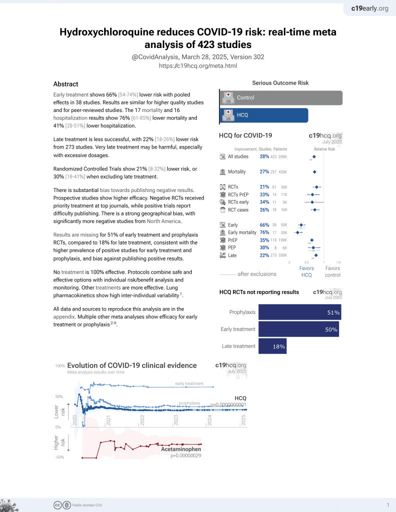
HCQ for COVID-19
1st treatment shown to reduce risk in
March 2020, now with p < 0.00000000001 from 424 studies, used in 59 countries.
No treatment is 100% effective. Protocols
combine treatments.
6,600+ studies for
220+ treatments. c19early.org
|

In vitro study presenting a new theory on SARS-CoV-2 infection and why HCQ/CQ provides benefits, which potentially explains the observed relationships with smoking, diabetes, obesity, age, and treatment delay, and confirms the importance of accurate dosing. Metabolic analysis revealed HCQ/CQ inhibit oxidative phosphorylation in mitochondria (likely by sequestering protons needed to drive ATP synthase), inhibiting infection and/or slowing replication.
40 preclinical studies support the efficacy of HCQ for COVID-19:
1.
Shang et al., Identification of Cathepsin L as the molecular target of hydroxychloroquine with chemical proteomics, Molecular & Cellular Proteomics, doi:10.1016/j.mcpro.2025.101314.
2.
González-Paz et al., Biophysical Analysis of Potential Inhibitors of SARS-CoV-2 Cell Recognition and Their Effect on Viral Dynamics in Different Cell Types: A Computational Prediction from In Vitro Experimental Data, ACS Omega, doi:10.1021/acsomega.3c06968.
3.
Alkafaas et al., A study on the effect of natural products against the transmission of B.1.1.529 Omicron, Virology Journal, doi:10.1186/s12985-023-02160-6.
4.
Guimarães Silva et al., Are Non-Structural Proteins From SARS-CoV-2 the Target of Hydroxychloroquine? An in Silico Study, ACTA MEDICA IRANICA, doi:10.18502/acta.v61i2.12533.
5.
Nguyen et al., The Potential of Ameliorating COVID-19 and Sequelae From Andrographis paniculata via Bioinformatics, Bioinformatics and Biology Insights, doi:10.1177/11779322221149622.
7.
Yadav et al., Repurposing the Combination Drug of Favipiravir, Hydroxychloroquine and Oseltamivir as a Potential Inhibitor Against SARS-CoV-2: A Computational Study, Research Square, doi:10.21203/rs.3.rs-628277/v1.
8.
Hussein et al., Molecular Docking Identification for the efficacy of Some Zinc Complexes with Chloroquine and Hydroxychloroquine against Main Protease of COVID-19, Journal of Molecular Structure, doi:10.1016/j.molstruc.2021.129979.
9.
Baildya et al., Inhibitory capacity of Chloroquine against SARS-COV-2 by effective binding with Angiotensin converting enzyme-2 receptor: An insight from molecular docking and MD-simulation studies, Journal of Molecular Structure, doi:10.1016/j.molstruc.2021.129891.
10.
Noureddine et al., Quantum chemical studies on molecular structure, AIM, ELF, RDG and antiviral activities of hybrid hydroxychloroquine in the treatment of COVID-19: molecular docking and DFT calculations, Journal of King Saud University - Science, doi:10.1016/j.jksus.2020.101334.
11.
Tarek et al., Pharmacokinetic Basis of the Hydroxychloroquine Response in COVID-19: Implications for Therapy and Prevention, European Journal of Drug Metabolism and Pharmacokinetics, doi:10.1007/s13318-020-00640-6.
12.
Rowland Yeo et al., Impact of Disease on Plasma and Lung Exposure of Chloroquine, Hydroxychloroquine and Azithromycin: Application of PBPK Modeling, Clinical Pharmacology & Therapeutics, doi:10.1002/cpt.1955.
13.
Pinatel et al., SARS-CoV-2 infects human primary cytotrophoblasts mainly through a non-canonical entry route, Molecular Human Reproduction, doi:10.1093/molehr/gaag015.
14.
Hitti et al., Hydroxychloroquine attenuates double-stranded RNA-stimulated hyper-phosphorylation of tristetraprolin/ZFP36 and AU-rich mRNA stabilization, Immunology, doi:10.1111/imm.13835.
15.
Yan et al., Super-resolution imaging reveals the mechanism of endosomal acidification inhibitors against SARS-CoV-2 infection, ChemBioChem, doi:10.1002/cbic.202400404.
16.
Mohd Abd Razak et al., In Vitro Anti-SARS-CoV-2 Activities of Curcumin and Selected Phenolic Compounds, Natural Product Communications, doi:10.1177/1934578X231188861.
17.
Alsmadi et al., The In Vitro, In Vivo, and PBPK Evaluation of a Novel Lung-Targeted Cardiac-Safe Hydroxychloroquine Inhalation Aerogel, AAPS PharmSciTech, doi:10.1208/s12249-023-02627-3.
18.
Wen et al., Cholinergic α7 nAChR signaling suppresses SARS-CoV-2 infection and inflammation in lung epithelial cells, Journal of Molecular Cell Biology, doi:10.1093/jmcb/mjad048.
19.
Kamga Kapchoup et al., In vitro effect of hydroxychloroquine on pluripotent stem cells and their cardiomyocytes derivatives, Frontiers in Pharmacology, doi:10.3389/fphar.2023.1128382.
20.
Milan Bonotto et al., Cathepsin inhibitors nitroxoline and its derivatives inhibit SARS-CoV-2 infection, Antiviral Research, doi:10.1016/j.antiviral.2023.105655.
21.
Miao et al., SIM imaging resolves endocytosis of SARS-CoV-2 spike RBD in living cells, Cell Chemical Biology, doi:10.1016/j.chembiol.2023.02.001.
22.
Yuan et al., Hydroxychloroquine blocks SARS-CoV-2 entry into the endocytic pathway in mammalian cell culture, Communications Biology, doi:10.1038/s42003-022-03841-8.
23.
Faísca et al., Enhanced In Vitro Antiviral Activity of Hydroxychloroquine Ionic Liquids against SARS-CoV-2, Pharmaceutics, doi:10.3390/pharmaceutics14040877.
24.
Delandre et al., Antiviral Activity of Repurposing Ivermectin against a Panel of 30 Clinical SARS-CoV-2 Strains Belonging to 14 Variants, Pharmaceuticals, doi:10.3390/ph15040445.
25.
Purwati et al., An in vitro study of dual drug combinations of anti-viral agents, antibiotics, and/or hydroxychloroquine against the SARS-CoV-2 virus isolated from hospitalized patients in Surabaya, Indonesia, PLOS One, doi:10.1371/journal.pone.0252302.
26.
Zhang et al., SARS-CoV-2 spike protein dictates syncytium-mediated lymphocyte elimination, Cell Death & Differentiation, doi:10.1038/s41418-021-00782-3.
27.
Dang et al., Structural basis of anti-SARS-CoV-2 activity of hydroxychloroquine: specific binding to NTD/CTD and disruption of LLPS of N protein, bioRxiv, doi:10.1101/2021.03.16.435741.
28.
Shang (B) et al., Inhibitors of endosomal acidification suppress SARS-CoV-2 replication and relieve viral pneumonia in hACE2 transgenic mice, Virology Journal, doi:10.1186/s12985-021-01515-1.
29.
Wang et al., Chloroquine and hydroxychloroquine as ACE2 blockers to inhibit viropexis of 2019-nCoV Spike pseudotyped virus, Phytomedicine, doi:10.1016/j.phymed.2020.153333.
30.
Sheaff, R., A New Model of SARS-CoV-2 Infection Based on (Hydroxy)Chloroquine Activity, bioRxiv, doi:10.1101/2020.08.02.232892.
31.
Ou et al., Hydroxychloroquine-mediated inhibition of SARS-CoV-2 entry is attenuated by TMPRSS2, PLOS Pathogens, doi:10.1371/journal.ppat.1009212.
32.
Andreani et al., In vitro testing of combined hydroxychloroquine and azithromycin on SARS-CoV-2 shows synergistic effect, Microbial Pathogenesis, doi:10.1016/j.micpath.2020.104228.
33.
Clementi et al., Combined Prophylactic and Therapeutic Use Maximizes Hydroxychloroquine Anti-SARS-CoV-2 Effects in vitro, Front. Microbiol., 10 July 2020, doi:10.3389/fmicb.2020.01704.
34.
Liu et al., Hydroxychloroquine, a less toxic derivative of chloroquine, is effective in inhibiting SARS-CoV-2 infection in vitro, Cell Discovery 6, 16 (2020), doi:10.1038/s41421-020-0156-0.
35.
Yao et al., In Vitro Antiviral Activity and Projection of Optimized Dosing Design of Hydroxychloroquine for the Treatment of Severe Acute Respiratory Syndrome Coronavirus 2 (SARS-CoV-2), Clin. Infect. Dis., 2020 Mar 9, doi:10.1093/cid/ciaa237.
Sheaff et al., 2 Aug 2020, preprint, 1 author.
In vitro studies are an important part of preclinical research, however results may be very different in vivo.
A New Model of SARS-CoV-2 Infection Based on (Hydroxy)Chloroquine Activity
doi:10.1101/2020.08.02.232892
Chloroquine and hydroxychloroquine (H)CQ are well known anti-malarial drugs, while their use against COVID-19 is more controversial. (H)CQ activity was examined in tissue culture cells to determine if their anti-viral benefits or adverse effects might be due to altering host cell pathways. Metabolic analysis revealed (H)CQ inhibit oxidative phosphorylation in mitochondria, likely by sequestering protons needed to drive ATP synthase. This activity could cause cardiotoxicity because heart muscle relies on beta oxidation of fatty acids. However, it might also explain their therapeutic benefit against COVID-19. A new model of SARS-CoV-2 infection postulates virus enters host cell mitochondria and uses its protons for genome release. Oxidative phosphorylation is eventually compromised, so glycolysis is upregulated to maintain ATP levels. (H)CQ could prevent viral infection and/or slow its replication by sequestering these protons. In support of this model other potential COVID-19 therapeutics also targeted mitochondria, as did tobacco smoke, which may underlie smokers' protection. The mitochondria of young people are naturally more adaptable and resilient, providing a rationale for their resistance to disease progression. Conversely, obesity and diabetes could exacerbate disease severity by providing extra glucose to infected cells dependent on glycolysis. The description of (H)CQ function presented here, together with its implications for understanding SARS-CO-V2 infection, makes testable predictions about disease progression and identifies new approaches for treating COVID-19. .
Smoke exposure Four p60mm plates of attached A549 lung carcinoma cells were aspirated, washed with PBS, and aspirated again. Control plates were untreated and exposed to breath exhalation in the absence of smoke. Cigar smoke was blown into two experimental plates followed by rapid lid closure to trap the smoke. After 3min. at room temperature the lid was removed from one plate and smoke allowed to dissipate (labeled 1 smoke in Figure 4G ). The other plate was exposed to another aliquot of cigar smoke followed by rapid lid closure (labeled 2 smoke). After 6 min. total incubation time cells were removed with trypsin, pelleted, and re-suspended in 500L of the indicated media. 100l aliquots were distributed in a white 96 well assay plate and incubated at 37 o C in the CO2 incubator for 1hr. ATP levels were then analyzed using CellTiterGlo as described previously.
Competing interests The author declares no competing interests.
Materials & Correspondence
Robert J. Sheaff Associate Professor The University of Tulsa Department of Chemistry and Biochemistry Keplinger M2215 800 South Tucker Drive Tulsa, OK 74104 Office 918-631-2319 E-mail: robert-sheaff@utulsa.edu Sheaff, 2020 Supl. Fig. 4 Supl. Fig. 5 : Red blood cells were pre-incubated with 1mM CQ for 1hr (orange line). After washing an aliquot was withdrawn to determine ATP levels using CTG (inset graph). Remaining cells were lysed by re-suspending in H2O and vortexing, followed by UV/Vis analysis of the..
References
Alifano, Alifano, Forgez, Iannelli, Renin-angiotensin system at the heart of COVID-19 pandemic, Biochimie, doi:10.1016/j.biochi.2020.04.008
Clayton, Shadel, Isolation of Mitochondria from Tissue Culture Cells, doi:10.1101/pdb.prot080002
Dowd, Andriano, Brazel, Rotondi, Block et al., Demographic science aids in understanding the spread and fatality rates of COVID-19, Proceedings of the National Academy of Sciences, doi:10.1073/pnas.2004911117
Felsenstein, Herbert, Mcnamara, Hedrich, COVID-19: Immunology and treatment options, Clin Immunol, doi:10.1016/j.clim.2020.108448
Ferner, Aronson, Chloroquine and hydroxychloroquine in covid-19, BMJ, doi:10.1136/bmj.m1432
Fitch, Involvement of heme in the antimalarial action of chloroquine, Trans Am Clin Climatol Assoc
Guzik, COVID-19 and the cardiovascular system: implications for risk assessment, diagnosis, and treatment options, Cardiovasc Res, doi:10.1093/cvr/cvaa106
Hussain, Bhowmik, Do, Moreira, COVID-19 and diabetes: Knowledge in progress, Diabetes Res Clin Pract, doi:10.1016/j.diabres.2020.108142
Janowitz, Gablenz, Pattinson, Wang, Conigliaro et al., Famotidine use and quantitative symptom tracking for COVID-19 in non-hospitalised patients: a case series, Gut, doi:10.1136/gutjnl-2020-321852
Kase, Nikolić, Bakke, Bogen, Aas et al., Remodeling of Oxidative Energy Metabolism by Galactose Improves Glucose Handling and Metabolic Switching in Human Skeletal Muscle Cells, PLoS One, doi:10.1371/journal.pone.0059972
Kashani, Hypoxia in COVID-19: Sign of Severity or Cause for Poor Outcomes, Mayo Clin Proc, doi:10.1016/j.mayocp.2020.04.021
Kolwicz, Purohit, Tian, Cardiac Metabolism and its Interactions with Contraction, Growth, and Survival of Cardiomyocytes, Circulation Research, doi:10.1161/CIRCRESAHA.113.302095
Magagnoli, Narendran, Pereira, Cummings, Hardin et al., Outcomes of hydroxychloroquine usage in United States veterans hospitalized with Covid-19, MedRxiv, doi:.10.1101/2020.04.16.20065920
Malik, Properties of Coronavirus and SARS-CoV-2, Malays J Pathol
Moore, Chloroquine for COVID-19 Infection, Drug Saf, doi:10.1007/s40264-020-00933-4
Osellame, Blacker, Duchen, Cellular and molecular mechanisms of mitochondrial function, Best Pract Res Clin Endocrinol Metab, doi:10.1016/j.beem.2012.05.003
Pajak, Siwiak, Sołtyka, Priebe, Zieliński et al., 2-Deoxy-d-Glucose and Its Analogs: From Diagnostic to Therapeutic Agents, Int J Mol Sci, doi:10.3390/ijms21010234
Park, Epidemiology, Virology, and Clinical Features of Severe Acute Respiratory Syndromecoronavirus-2 (SARS-CoV-2
Remington, Powell, Clozapine and COVID-19, J Psychiatry Neurosci, doi:10.1503/jpn.2045301
Schrezenmeier, Dörner, Mechanisms of action of hydroxychloroquine and chloroquine: implications for rheumatology, Nat Rev Rheumatol, doi:10.1038/s41584-020-0372-x
Slater, Chloroquine: mechanism of drug action and resistance in Plasmodium falciparum, Pharmacol Ther, doi:10.1016/0163-7258(93)90056-j
Sun, Youle, Finkel, The Mitochondrial Basis of Aging, Mol Cell, doi:10.1016/j.molcel.2016.01.028
Vardavas, Nikitara, COVID-19 and smoking: A systematic review of the evidence, Tob Induc Dis, doi:10.18332/tid/119324
Vial, Detaille, Guigas, Role of Mitochondria in the Mechanism(s) of Action of Metformin, Front Endocrinol, doi:10.3389/fendo.2019.00294
Walls, Park, Tortorici, Wall, Mcguire et al., Structure, Function, and Antigenicity of the SARS-CoV-2 Glycoprotein, Cell, doi:10.1016/j.cell.2020.02.058
Weiss, Leibowitz, Coronavirus pathogenesis, Adv Virus Res, doi:10.1016/B978-0-12-385885-6.00009-2
Xue, Moyer, Peng, Wu, Hannafon et al., Chloroquine is a zinc ionophore, PLoS One, doi:10.1371/journal.pone.0109180
DOI record:
{
"DOI": "10.1101/2020.08.02.232892",
"URL": "http://dx.doi.org/10.1101/2020.08.02.232892",
"abstract": "<jats:p>Chloroquine and hydroxychloroquine [(H)CQ] are well known anti-malarial drugs, while their use against COVID-19 is more controversial. (H)CQ activity was examined in tissue culture cells to determine if their anti-viral benefits or adverse effects might be due to altering host cell pathways. Metabolic analysis revealed (H)CQ inhibit oxidative phosphorylation in mitochondria, likely by sequestering protons needed to drive ATP synthase. This activity could cause cardiotoxicity because heart muscle relies on beta oxidation of fatty acids. However, it might also explain their therapeutic benefit against COVID-19. A new model of SARS-CoV-2 infection postulates virus enters host cell mitochondria and uses its protons for genome release. Oxidative phosphorylation is eventually compromised, so glycolysis is upregulated to maintain ATP levels. (H)CQ could prevent viral infection and/or slow its replication by sequestering these protons. In support of this model other potential COVID-19 therapeutics also targeted mitochondria, as did tobacco smoke, which may underlie smokers protection. The mitochondria of young people are naturally more adaptable and resilient, providing a rationale for their resistance to disease progression. Conversely, obesity and diabetes could exacerbate disease severity by providing extra glucose to infected cells dependent on glycolysis. The description of (H)CQ function presented here, together with its implications for understanding SARS-CO-V2 infection, makes testable predictions about disease progression and identifies new approaches for treating COVID-19.</jats:p>",
"accepted": {
"date-parts": [
[
2020,
8,
2
]
]
},
"author": [
{
"affiliation": [],
"family": "Sheaff",
"given": "Robert J.",
"sequence": "first"
}
],
"container-title": [],
"content-domain": {
"crossmark-restriction": false,
"domain": []
},
"created": {
"date-parts": [
[
2020,
8,
3
]
],
"date-time": "2020-08-03T08:45:15Z",
"timestamp": 1596444315000
},
"deposited": {
"date-parts": [
[
2020,
8,
3
]
],
"date-time": "2020-08-03T08:45:15Z",
"timestamp": 1596444315000
},
"group-title": "Cell Biology",
"indexed": {
"date-parts": [
[
2023,
7,
5
]
],
"date-time": "2023-07-05T09:30:54Z",
"timestamp": 1688549454132
},
"institution": [
{
"name": "bioRxiv"
}
],
"is-referenced-by-count": 1,
"issued": {
"date-parts": [
[
2020,
8,
2
]
]
},
"link": [
{
"URL": "https://syndication.highwire.org/content/doi/10.1101/2020.08.02.232892",
"content-type": "unspecified",
"content-version": "vor",
"intended-application": "similarity-checking"
}
],
"member": "246",
"original-title": [],
"posted": {
"date-parts": [
[
2020,
8,
2
]
]
},
"prefix": "10.1101",
"published": {
"date-parts": [
[
2020,
8,
2
]
]
},
"publisher": "Cold Spring Harbor Laboratory",
"reference-count": 0,
"references-count": 0,
"relation": {},
"resource": {
"primary": {
"URL": "http://biorxiv.org/lookup/doi/10.1101/2020.08.02.232892"
}
},
"score": 1,
"short-title": [],
"source": "Crossref",
"subject": [],
"subtitle": [],
"subtype": "preprint",
"title": "A New Model of SARS-CoV-2 Infection Based on (Hydroxy)Chloroquine Activity",
"type": "posted-content"
}